Touch sensing skin can interface directly to nerves/brain..

flacaltenn

Diamond Member
Jun 9, 2011
67,573
22,953
2,250
Hillbilly Hollywood, Tenn
This is not just for robotics -- but has large promise in prosthetic devices.. The bigger part here is discovering the signal coding and communications for a touch as seen by the nerve cells and then it becomes possible to transmit that to the brain as a seamless replacement for lost limbs/fingers/toes..


Stanford engineers create artificial skin that can send pressure sensation to brain cell

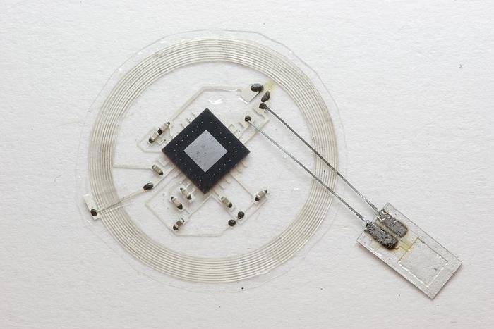
Stanford engineers have created a plastic "skin" that can detect how hard it is being pressed and generate an electric signal to deliver this sensory input directly to a living brain cell.
Shenan Bao, a professor of chemical engineering at Stanford, has spent a decade trying to develop a material that mimics skin's ability to flex and heal, while also serving as the sensor net that sends touch, temperature and pain signals to the brain. Ultimately she wants to create a flexible electronic fabric embedded with sensors that could cover a prosthetic limb and replicate some of skin's sensory functions.

Bao's work, reported today in Science, takes another step toward her goal by replicating one aspect of touch, the sensory mechanism that enables us to distinguish the pressure difference between a limp handshake and a firm grip. See video here.

"This is the first time a flexible, skin-like material has been able to detect pressure and also transmit a signal to a component of the nervous system," said Bao, who led the 17-person research team responsible for the achievement.

Benjamin Tee, a recent doctoral graduate in electrical engineering; Alex Chortos, a doctoral candidate in materials science and engineering; and Andre Berndt, a postdoctoral scholar in bioengineering, were the lead authors on the Science paper.
 
upload_2015-10-25_22-27-31.jpeg


Soon you'll all be part of the collective.

*****CHUCKLE*****


:)
 
Seeking Ways to Help Heal Damaged Brains...

Researchers Seek Ways to Help Damaged Brains Heal
December 15, 2015 — Using powerful microscopes and imaging devices, Rice University researcher Amina Qutub can observe brain cells in a glass container and the connections they make with each other.
Normal brain activity results from those cells — called neurons — linking together in complex networks, and researchers are seeking a better understanding of how that happens. "We are looking at how neural progenitors, which are cells that can regenerate in the brain, form active neural networks," she said. Brain cells connect with each other through both chemical and electrical signals, and they utilize a variety of proteins to stimulate growth. Qutub's team of Rice University scientists and technicians combine such fields as biology, electrical engineering and nanotechnology to unravel the mysteries of how this works. "Any one cell here is influenced by all of its neighbors in the environment,” Qutub said. “So, we are teasing out how that one cell, as it becomes an electrically active neuron, is affected by the community of cells."

‘Elaborate, beautiful structures’

In the laboratory there is a piece of abstract art that is composed of images of neuron networks, showing their vast complexity. "They form very elaborate and beautiful structures,” Qutub said of the neurons, “and the different network structures relate to what they do, their function. What we are trying to figure out is what are the chemical signals that lead to a particular structure and, in turn, how does that chemical structure lead to the electrical signals that you get when you have active neurons." The Rice research is supported in part by President Barack Obama's $300 million BRAIN Initiative, which is modeled after the Human Genome Project.

The research “opens up a huge door to better understanding the brain," Qutub said. Like many other people around the world, Qutub has seen how a stroke or a disease such as Alzheimer's can disrupt brain function, and she hopes this research will one day provide better treatment. "We are understanding the mechanism of how these cells can grow back, and with it we can identify better drugs and better targets from the basic science work," she said. For now, the research team is focused on developing a dynamic computer model that can help analyze interactions in the world's most complex mechanism.

Video
 
New implantable brain monitor dissolves when no longer needed...

Implantable brain monitor dissolves when no longer needed
Jan. 18, 2016 - The sensors may also be built into similar monitors that can be used for other organs.
Researchers created thin, electronic sensors that can be implanted in the brain after injury to monitor temperature and pressure, and then dissolve when they are no longer needed. The sensors, developed by scientists at the University of Illinois and Washington University School of Medicine in St. Louis, may also be built into similar monitors that can be used for other organs.

Although there are many devices used to monitor activity in the body, they can be large, unwieldy and sometimes require surgery to be removed. Those that are implanted also often carry the risk of infection or rejection by the body, which the scientists said is avoided because the new devices dissolve away. "The ultimate strategy is to have a device that you can place in the brain -- or in other organs in the body -- that is entirely implanted, intimately connected with the organ you want to monitor and can transmit signals wirelessly to provide information on the health of that organ, allowing doctors to intervene if necessary to prevent bigger problems," said Dr. Rory Murphy, a neurosurgery resident at Washington University School of Medicine, in a press release. "And then after the critical period that you actually want to monitor, it will dissolve away and disappear."

Implantable-brain-monitor-dissolves-when-no-longer-needed.jpg

The small sensor connects to an embeddable wireless transmitter that lies on top of the skull.​

In a study published in the journal Nature, the researchers describe the devices, which are made out of polylactic-co-glycolic acid and silicone. The devices were first tested in saline, which caused them to dissolve within a few days. Researchers then tested them in rats, taking accurate measurements and then seeing them dissolve successfully. The researchers said the next step is to start testing the devices in humans for both safety and accuracy. "With advanced materials and device designs, we demonstrated that it is possible to create electronic implants that offer high performance and clinically relevant operation in hardware that completely resorbs into the body after the relevant functions are no longer needed," said Dr. John Rogers, a professor of materials science and engineering at the University of Illinois. "This type of bio-electric medicine has great potential in many areas of clinical care."

Implantable brain monitor dissolves when no longer needed

See also:

Most younger people with stroke symptoms would not go to ER
Jan. 11, 2016 - Nearly three-quarters of people under age 45 would wait to see if symptoms subsided or got worse before seeking treatment.
People under age 45 may underestimate or ignore stroke symptoms and delay going to the hospital for treatment, according to a new survey of Americans. Researchers at the University of California Los Angeles were surprised to find nearly three-quarters of people they spoke with would wait to see if the symptoms subsided or got worse. Treatment for a stroke is critical within the first few hours in order to minimize or reverse damage to the brain. Stroke is the fifth leading cause of death in the United States, according to the Centers for Disease Control and Prevention, killing about 130,000 people per year. There are approximately 795,000 strokes in the United States each year, about 610,000 of which are first or new strokes, the agency reports.

Most-younger-people-with-stroke-symptoms-would-not-go-to-ER.jpg

Dr. David Liebeskind examines Jennifer Reilly, who had a stroke at age 27. Reilly waited weeks to seek treatment while periodically experiencing numbness in one of her hands before waking up in the middle of the night with an excruciating headache -- and didn't go to the hospital until a coworker insisted the next day.​

Previous research also has shown there has been up to a 53 percent increase in the number of strokes experienced by Americans between the ages of 15 and 44. "Timely treatment for stroke is probably more important than for almost any other medical problem there is," said Dr. David Liebeskind, a professor of neurology at Ronald Reagan UCLA Medical Center, in a press release. "There is a very limited window in which to start treatment because the brain is very sensitive to a lack of blood flow or to bleeding, and the longer patients wait, the more devastating the consequences." Researchers at UCLA interviewed more than 1,000 people about their actions within the first three hours of experiencing common symptoms of stroke such as weakness, numbness, difficulty speaking, or difficulty seeing.

Just one out of three who participated in the survey said they would be "very likely" to go to the hospital, while 73 percent of participants said they would wait to see if symptoms improved or got worse before seeking treatment. Researchers said part of this is a lack of familiarity with the symptoms or their potential seriousness. In addition to understanding risk factors and lifestyle decisions that can lower the risk of stroke, the researchers suggest keeping in mind the acronym F.A.S.T., which stands for: "Face drooping, Arm weakness, or Speech difficulty -- it's Time to call 911." "Believe it or not, it's on the order of minutes or hours when somebody has to seek medical attention," Liebeskind said. "There simply is no time to wait. It's a message that we clearly need to get to younger people more effectively."

Most younger people with stroke symptoms would not go to ER

Related:

Lights, music in casinos interact with brain like drug addiction
Jan. 20, 2016 -- Flashing lights and music encourage rats to make risky decisions in a "rat casino" in ways similar to their effect on humans, which scientists said may offer some explanation for gambling addiction.
Scientists at the University of British Columbia found rats were more likely to engage in risky, gambling-like behavior with bright lights and loud sounds -- and were less likely when a specific dopamine receptor was blocked in their brain.

The dopamine D3 receptor is already suspected to be important to drug addiction, meaning the new study supports theories that addictions have a common biological cause. "Anyone who's ever designed a casino game or played a gambling game will tell you that of course sound and light cues keep you more engaged, but now we can show it scientifically," said Dr. Catharine Winstanley, an associate professor in the Department of Psychology at the University of British Columbia, in a press release. "I often feel that scientific models are decades behind the casinos. I don't think it's an accident that casinos are filled with lights and noise."

In the study, published in the journal Neuroscience, researchers trained rats to play gambling-like games. The rats then had to choose between four reward and punishment gambling options and they were tested for their response with and without lights and loud sounds.

While the scientists report rats generally learn to avoid risky behaviors that result in punishment, the light and sound caused them to continue taking larger risks. When the scientists administered a drug that blocked the dopamine D3 receptor, the rats' risky decision-making decreased. "This brain receptor is also really important to drug addiction, so our findings help support the idea that risky behavior across different vices might have a common biological cause," said Michael Barrus, a doctoral candidate at the University of British Columbia.

Lights, music in casinos interact with brain like drug addiction
 
Last edited:

Forum List

Back
Top