wearable EEG isolates frontal midline theta

scruffy

Diamond Member
Joined
Mar 9, 2022
Messages
30,089
Reaction score
26,748
Points
2,788
Frontal midline theta is a brain wave that occurs when you concentrate intently. When you're firing a rifle or solving a math problem you get lots of frontal midline theta.

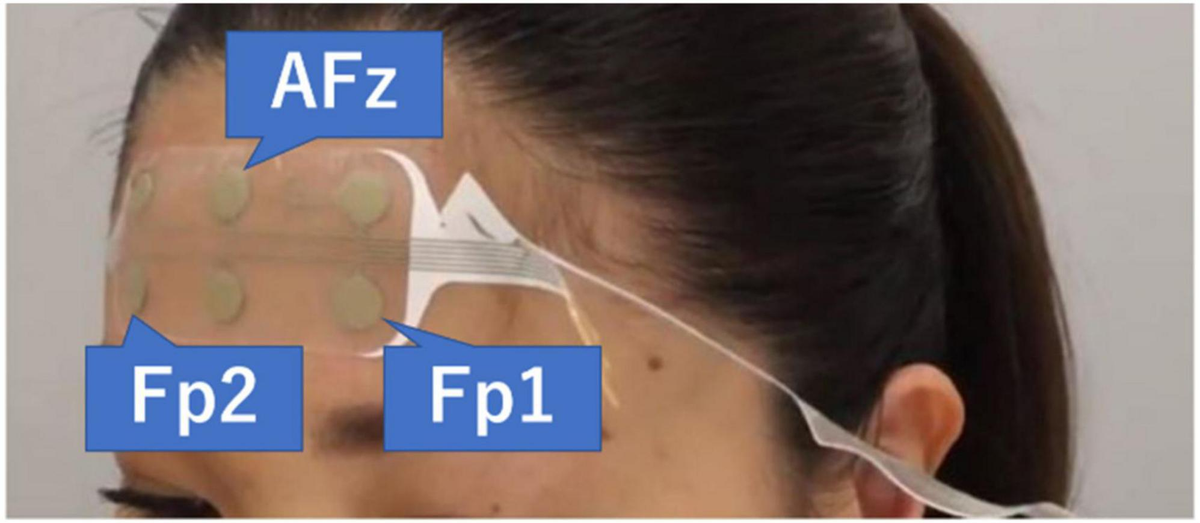
Researchers are now measuring this using wearable fabric-type EEG electrodes. They don't need gel, basically you just slap them on the forehead and they stay there.

1769405406365.webp


The wire connects to a little Bluetooth enabled belt pack so you can look at the signal on your cell phone.


This is very cool. This is called "synthetic aperture magnetometry".

What they do here is combine the broad and diffuse EEG with an MEG "beam former" that precisely localizes current sources.

This is showing you the location of the frontal midline theta generator.

1769420255902.webp


As you can see, it moves around.

So we have this other phenomenon, where the 6.5 Hz theta couples to both alpha (11 Hz) and gamma (22-30 Hz) in different areas of the brain. When frontal midline theta is active, the default mode network shuts down. The frontal lobe takes over and starts running the show.

The Kuramoto model shows that the time constants are much too long for any phase coupling during the few milliseconds between neighboring theta peaks. Synchronization has to happen externally, and it does, and we know exactly how it happens.

Underlying all this, is brain wiring called a syncytium, which is basically a subpopulation of the neural network connected by electrically conductive gap junctions. They're like little ohmic resistors, and their time constants are in the microseconds, rather than the milliseconds of chemical synapses. Local oscillators are coupled through an inhibitory syncytium, which also serves to synchronize them. Spikes ride on top of all this, they happen during the peaks of theta.

With current source density mapping you can see the shapes of the generators in four dimensions, like this:

1769421706722.webp


The time axis is along the bottom. In this particular case we're coupling to 90 msec which is about 11 Hz which is alpha. On the vertical axis are the layers in a different part of the brain near the hippocampus.

This is a great set of tools. The EEG patches in the OP are completely non magnetic, so you can put them on then put the subject in an MRI or an MEG with no ill effects.
 
The second brain image from the left, on the bottom, is especially interesting.

It's showing a source in the anterior cingulate cortex.

This proves that the focus of theta moves around quite considerably, not only within the frontal lobe, but even outside it, to areas that are connected with it yet distant.

There is an entirely separate theta wave system in the hippocampus and dorsolateral prefrontal cortex. It's the one that everyone knows about, that does the place cells and etc. The phase encoding mechanism has been well studied by now.

There's another one in the parietal lobe, it's connected to visual area V4. So this pattern seems to be ubiquitous, it's repeated many times. The question is, how does it enable memory, because each of these areas has a short term or working memory - and it's one-shot, it only requires one presentation.
 

New Topics

Back
Top Bottom