Brain & mental disorder research

ECT 'turns down' brain connection...
:confused:
Electroconvulsive therapy: Aberdeen team says it 'turns down overactive connection'
19 March 2012 - ECT has been used as a controversial treatment since the 1930s
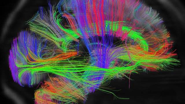
Electroconvulsive therapy for the severely depressed works by "turning down" an overactive connection between areas of the brain, Aberdeen researchers have claimed. ECT - which involves placing electrodes on the temples and delivering a small electrical current - has been used by psychiatrists since the 1930s. The aim is to shock the brain and to restore its natural chemical balance. The University of Aberdeen team hopes the findings can help treatment.

In a paper published in the journal, Proceedings of the National Academy of Sciences (PNAS), the researchers have said that the treatment appears to turn down an overactive connection between areas of the brain that control mood and the parts responsible for thinking and concentrating. This, they argue, stops the impact that depression has on the ability to enjoy life. The study involved using MRI scans of the brains of nine severely depressed patients before and after ECT.

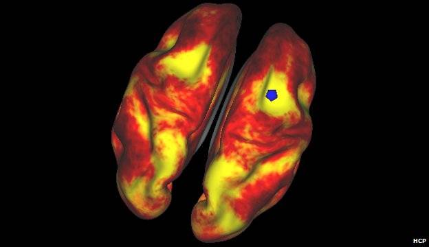
Prof Ian Reid, who led the research, said: "ECT is a controversial treatment, and one prominent criticism has been that it is not understood how it works and what it does to the brain. "However we believe we've solved a 70-year-old therapeutic riddle because our study reveals that ECT affects the way different parts of the brain involved in depression connect with one another. "For all the debate surrounding ECT, it is one of the most effective treatments, not just in psychiatry, but in the whole of medicine, because 75% to 85% of patients recover from the symptoms." He added: "Over the last couple of years there has been an emerging new perspective on how depression affects the brain.

"This theory has suggested a 'hyperconnection' between the areas of the brain involved in emotional processing and mood change and the parts of the brain involved in thinking and concentrating. "Our key finding is that if you compare the connections in the brain before and after ECT, ECT reduces the connection strength between these same areas - it reduces this hyperconnectivity. "For the first time we can point to something that ECT does in the brain that makes sense in the context of what we think is wrong in people who are depressed." The researchers now hope to continue monitoring the patients to see if the depression and hyperconnectivity returns.

'Make sense'
 
Granny always wears her old metal army helmet when she goes to the dentist...
:eusa_shifty:
Study Links Dental X-Rays to Brain Tumors
April 16, 2012 - Risk doubles compared to common x-rays
A new study links dental x-rays to the most common kind of brain tumor. But the risk of these mostly non-cancerous growths is still very small, even if your dentist takes an aggressive approach to imaging. For the study published in Cancer, about 1,400 patients - who had had the brain tumors called meningiomas - were interviewed about their family's health, their own medical history, exposure to radiation, and so on. They were compared with a control group that didn't have brain tumors.

Dr. Elizabeth Claus of Yale University, who led the study, says there was a clear relationship between dental x-rays and the brain tumors. "We found that the individuals that were diagnosed with meningioma were essentially twice as likely to report having a specific type of x-ray, which is very common, a bitewing." Bitewing x-rays are the ones where you clamp down - usually with your back teeth - on a holder for film or a sensor. The link between these kinds of brain tumors and a different kind of imaging, called a panoramic x-ray, was even stronger - up to five times the risk, depending on age or how frequently the x-ray was taken.

All studies have strengths and weaknesses, and Claus says one issue was that the people in this study - typically in their 50s or older - were being asked about x-rays they might have had decades earlier. "There is always some difficulty in people remembering past exposures. And so patients could under- or over-report their dental x-rays. So you always have to keep that in mind." That applies to both groups, of course - the meningioma patients and the control group.

Today, dentists are more aware that, as useful as x-rays are, they can also cause harm. Maybe your dentist covers you with a lead apron when you get an x-ray. And modern x-ray equipment delivers a much lower dose than was common in years past. So Claus doesn't want to scare people away from regular dental check-ups. "The big take-home message is, keep going to the dentist," she says. "But have a conversation with your dentist about whether you might be able to reduce the number of x-rays that you receive."

Source
 
Control seizures & lose weight with the same drug...
:cool:
'Fat' drug could treat epilepsy
21 November 2012 - A substance made by the body when it uses fat as fuel could provide a new way of treating epilepsy, experts hope.
Researchers in London who have been carrying out preliminary tests of the fatty acid treatment, report their findings in Neuropharmacology journal. They came up with the idea because of a special diet used by some children with severe, drug resistant epilepsy to help manage their condition. The ketogenic diet is high in fat and low in carbohydrate. The high fat, low carbohydrate diet is thought to mimic aspects of starvation by forcing the body to burn fats rather than carbohydrates. Although often effective, the diet has attracted criticism, as side-effects can be significant and potentially lead to constipation, hypoglycaemia, retarded growth and bone fractures. By pinpointing fatty acids in the ketogenic diet that are effective in controlling epilepsy, researchers hope they can develop a pill for children and adults that could provide similar epilepsy control without the side-effects.

In early trials, the scientists, from Royal Holloway and University College London, say they have identified fatty acids that look like good candidates for the job. They found that not only did some of the fatty acids outperform a regular epilepsy medication called valproate in controlling seizures in animals, they also had fewer side-effects. But many more tests are needed to determine if the treatment would be safe and effective in humans. Prof Matthew Walker, from the Institute of Neurology, University College London, said: "Epilepsy affects over 50 million people worldwide and approximately a third of these people have epilepsy that is not adequately controlled by our present treatments. "This discovery offers a whole new approach to the treatment of drug-resistant epilepsies in children and adults."

Simon Wigglesworth, deputy chief executive at Epilepsy Action, said: "We know the ketogenic diet can be a highly effective treatment for children with difficult to control epilepsy and it is starting to be used for adults. "The diet is high in fats and low in carbohydrates and the balance of the diet needs to be carefully worked out for each child. Although some children manage the diet very well, others find the diet unpleasant and difficult to follow. Children can also experience side-effects including constipation and weight loss. "The identification of these fatty acids is an exciting breakthrough. The research means that children and adults with epilepsy could potentially benefit from the science behind the ketogenic diet without dramatically altering their eating habits or experiencing unpleasant side-effects. "We look forward to seeing how this research progresses."

BBC News - 'Fat' drug could treat epilepsy
 
Researchers say they found a strong genetic basis for migraine and epilepsy...
:cool:
Epilepsy and migraine 'could have shared genetic link'
7 January 2013 - A strong family history of seizures could increase the chances of having severe migraines, says a study in Epilepsia journal.
Scientists from Columbia University, New York, analysed 500 families containing two or more close relatives with epilepsy. Their findings could mean that genes exist that cause both epilepsy and migraine. Epilepsy Action said it could lead to targeted treatments. Previous studies have shown that people with epilepsy are substantially more likely than the general population to have migraine headaches, but it was not clear whether that was due to a shared genetic cause.

The researchers found that people with three or more close relatives with a seizure disorder were more than twice as likely to experience 'migraine with aura' than patients from families with fewer individuals with seizures. Migraine with aura is a severe headache preceded by symptoms such as seeing flashing lights, temporary visual loss, speech problems or numbness of the face.

Dr Melodie Winawer, lead author of the study from Columbia University Medical Centre, said the findings had implications for epilepsy patients. "Our study demonstrates a strong genetic basis for migraine and epilepsy, because the rate of migraine is increased only in people who have close (rather than distant) relatives with epilepsy." She said further studies of disorders coupled with epilepsy could help "enhance the quality of life for those with epilepsy". In the study, Dr Winawer and colleagues analysed data collected from the Epilepsy Phenome/Genome Project - a genetic study of epilepsy patients and families from 27 clinical centres in the US, Canada, Argentina, Australia, and New Zealand.

Simon Wigglesworth, deputy chief executive at Epilepsy Action, said the research was encouraging. "Having a better understanding of the genetic link between epilepsy and other medical conditions can only be a good thing. "It means that steps can be taken to improve diagnosis and treatment for people living epilepsy and co-existing conditions. By understanding how genes work, more targeted treatments could be developed in the future."

BBC News - Epilepsy and migraine 'could have shared genetic link'
 
Childhood epilepsy in steep decline...
:clap2:
'Steep decline' in child epilepsy
2 February 2013 - The number of children being diagnosed with epilepsy has dropped dramatically in the UK over the past decade, figures show.
A study of GP-recorded diagnoses show the incidence has fallen by as much as half. Researchers said fewer children were being misdiagnosed, but there had also been a real decrease in some causes of the condition. Other European countries and the US had reported similar declines, they added. Epilepsy is caused when the brain's normal electrical activity result in seizures. Data from more than 344,000 children showed that the annual incidence of epilepsy has fallen by 4-9% year on year between 1994 and 2008. Overall the number of children born between 2003-2005 with epilepsy was 33% lower then those born in 1994-96. When researchers looked in more detail and included a wider range of possible indicators of an epilepsy diagnosis the incidence dropped by 47%.

Correct diagnosis

Better use of specialist services and increased caution over diagnosing the condition explains some, but not all, of the decline in the condition, the researchers reported in Archives of Diseases in Childhood. Introduction of vaccines against meningitis and a drop in the number of children with traumatic brain injuries, both of which can cause epilepsy, has probably also contributed to falling cases, they added.

Study author Prof Ruth Gilbert, director of the Centre for Evidence-based Child Health at University College London, said: "The drop is consistent with what has been seen in other countries so it is reassuring that we are seeing the same pattern. "We're getting better at diagnosing and deciding who should be treated and then there is also probably an effect of factors like fewer cases of meningitis." She said in the past, there was an issue with variable diagnosis and some children being treated who did not need to be. "There is a more rigorous approach and that is partly down to NICE guidance. "It is very troubling to have a misdiagnosis because once you have a diagnosis it sticks and that does blight the life of a child."

Simon Wigglesworth, deputy chief executive at Epilepsy Action, said: "It may indicate a reduction in misdiagnosis rates in children, which we know to be high. However, our discussions with leading clinicians suggest that this may not be the complete picture. "They tell us that they are not seeing a reduction in the number of children with epilepsy presenting at their clinics and epilepsy remains one of the most prevalent neurological conditions in children in the UK."

BBC News - 'Steep decline' in child epilepsy
 
Uncle Ferd says dey oughta map Granny's brain so she don't get lost...
:redface:
Scans reveal intricate brain wiring
16 February 2013 - Scientists are set to release the first batch of data from a project designed to create the first map of the human brain.
The project could help shed light on why some people are naturally scientific, musical or artistic. Some of the first images were shown at the American Association for the Advancement of Science meeting in Boston. I found out how researchers are developing new brain imaging techniques for the project by having my own brain scanned. Scientists at Massachusetts General Hospital are pushing brain imaging to its limit using a purpose built scanner. It is one of the most powerful scanners in the world.

_65931406_65931405.jpg

The BBC's Pallab Ghosh gets a look at how his brain is wired

The scanner's magnets need 22MW of electricity - enough to power a nuclear submarine. The researchers invited me to have my brain scanned. I was asked if I wanted "the 10-minute job or the 45-minute 'full monty'" which would give one of the most detailed scans of the brain ever carried out. Only 50 such scans have ever been done. I went for the full monty.

It was a pleasant experience enclosed in the scanner's vast twin magnets. Powerful and rapidly changing magnetic fields were looking to see tiny particles of water travelling along the larger nerve fibres.By following the droplets, the scientists in the adjoining cubicle are able to trace the major connections within my brain.

Arcs of understanding
 
Brain has partial ability to shield itself from the destructive damage caused by a stroke...
:cool:
Brain's 'stroke shielding' cracked
24 February 2013 - A part of the brain's ability to shield itself from the destructive damage caused by a stroke has been explained by researchers.
It has been known for more than 85 years that some brain cells could withstand being starved of oxygen. Scientists, writing in the journal Nature Medicine, have shown how these cells switch into survival mode. They hope to one-day find a drug which uses the same trick to protect the whole brain. Treating a stroke is a race against time. Clots that block the blood supply prevent the flow of oxygen and sugar to brain cells, which then rapidly die. But in 1926, it was noticed that some cells in the hippocampus, the part of the brain involved in memory, did not follow this rule. "They're staying alive when the prediction would say that they should die," said Prof Alastair Buchan from Oxford University who has investigated how they survive.

I'm a survivor

Experiments on rats showed that these surviving-cells started producing a protein called hamartin - which forces cells to conserve energy. They stop producing new proteins and break down existing ones to access the raw materials. When the researchers prevented the cells from producing hamartin, they died just like other cells. Prof Buchan said: "We have shown for the first time that the brain has mechanisms that it can use to protect itself and keep brain cells alive."

_66020802_c0119505-stroke,_conceptual_artwork-spl.jpg


Their aim is to develop a drug that can produce the same effect, which could be given when an ambulance arrived. This would buy the brain time until clot-busting drugs could be given in hospital. The researchers do not know why these cells have this defence, but other nearby cells in the hippocampus do not. There are differences in function. The cells that die are known as CA1 cells which are very plastic and are involved in laying down memories whereas the surviving, or CA3, cells are less adaptable. Speaking to BBC News online, Prof Buchan said the focus of this research was on "ways to keep brain cells alive" which could have impacts beyond stroke - such as in Alzheimer's disease and spinal cord injuries.

Commenting on the study, Dr Clare Walton from the Stroke Association said: "Previous research has shown that some brain cells are naturally more resilient than others, and this study has identified a particular protein in the cells that is responsible. "In the future, researchers could try to turn on this protein in other, less resilient brain cells to reduce the brain damage caused by stroke. "The findings of this research are exciting, but we are still a long way off from developing a new stroke treatment."

BBC News - Brain's 'stroke shielding' cracked
 
Human Connectome Project scans will reveal how brain works...
:cool:
World's most detailed scans will reveal how brain works
5 March 2013 - Scientists say they have published the most detailed brain scans "the world has ever seen" as part of a project to understand how the organ works.
The aim of the project is to determine how a person's brain structure influences their talents and behaviour. Researchers involved in the so called Human Connectome Project have published the scans of 68 adults in the study. They eventually hope to scan 1,200 people and also collect details of their behavioural traits and DNA. The information is made freely available to neuroscientists in their quest to unlock the secrets of the human brain. The project leader, Prof David Van Essen of Washington University in St Louis, told BBC News that sharing the data with the international community of researchers would spur rapid advances in brain science. "We are very optimistic that as the community delves in and begins working on these data sets, they will reveal new insights into the brain circuits of healthy adults," he said.

_66196102_full_correl_all_infl.jpg

Daydream Believer: an MRI scan of the brain at rest. Regions in yellow are strongly linked to the area indicated by the blue spot.

Subjects involved in the project have their brain scanned for a total of four hours. For part of that time, they carry out a battery of tasks, which include arithmetic, listening to stories, gambling and moving parts of their body. Volunteers also engage in tests that assess their skills and abilities. In addition, DNA samples are taken. The scans are essentially a wiring diagram for each person's brain. They show how different parts are connected by nerve fibres and also the thickness of the bundles, which is thought to be an indication of the importance or strength of a particular connection- a so-called "structural map". Scanning can also show which parts of the brain are activated for particular tasks - known as a "functional map". With all this information, researchers will be able to see if an individual's brain wiring is related to their skills, such as musicality, sociability and aptitude for science or maths.

Neural circuitry

According to Oxford University's Dr Tim Behrens, who is collaborating with Prof Van Essen, the study will "uncover which neural pathways are important in determining human behaviours". The eventual aim of the project is to understand how the healthy human brain is wired and how differences between individuals make each person unique - shaping their personalities and their capacity to think and feel. "We have the highest quality data of the entire human brain that the world has ever seen. The question is that with more cutting edge (scanning) methods, how much can we decipher the circuits that give us our distinctive capabilities?" he said.

By learning more about how the healthy human brain works, the research will inevitably be of use to those studying brain disorders, such as Alzheimer's and Parkinson's. Among those who will be delving through the data is Ed Bullmore, a professor of psychiatry at Cambridge University. He believes that psychiatric problems, such as schizophrenia, drug addiction and obsessive compulsive disorder are generally arise from irregular brain development.

Dementia
 
Brain implant could be epilepsy early seizure warning system...
:cool:
Brain implant 'predicts' epilepsy seizures
1 May 2013 - A brain implant may be able to predict epilepsy seizures by picking up the early warning signs, a small study suggests.
The device uses the brain's electrical activity to tell patients if their risk of a seizure is high, moderate or low. The study on 15 people, published in the Lancet Neurology, showed the device worked in some patients. The charity Epilepsy Action cautioned that it was still early days, but said it could be an "exciting development". Epilepsy is thought to affect 50 million people worldwide. Abnormal activity in part of the brain causes seizures involving involuntary shaking.

Independence impact

Signals were collected from the surface of the brain and sent down wires to another implant in the chest. This beamed the data to a hand-held device which worked out the odds of a seizure. The trial was run at three hospitals in Australia and was funded by the manufacturers NeuroVista. The results were mixed. For the first four months the brain was monitored so the system could learn a patient's brainwaves before a seizure. Only eight patients then progressed to the stage where the device was fully activated and they were constantly informed of their chance of a seizure. It was between 56% and 100% effective in those patients.

Prof Mark Cook, from the University of Melbourne, said if the technology could be proven if could help remove the unpredictable nature of epilepsy. He told the BBC: "Being able to predict the events with many minutes or hours lead time could have significant impact on independence. "This could change the way the illness is treated. For instance, our current strategy of giving medications continuously because of the unpredictable occurrence of events could alter the types of medications being developed. "Short-acting therapies may prove to be effective without subjecting patients to the long-term problems that currently available therapies may cause."

'Useful tool'

Commenting on the findings, Christian Elger and Florian Mormann, from the University of Bonn medical centre, described the results as "a major milestone... showing for the first time, to our knowledge that prospective seizure prediction is possible". They added: "Whether this performance is also sufficient for clinical applications is unclear, this will depend on how well patients tolerate false alarms or missed seizures."

Simon Wigglesworth, deputy chief executive of Epilepsy Action, said more research was needed, particularly given the "small sample size and the inconsistencies in the data collected". "If a person is able to be alerted when they are about to have a seizure, this could help them to take steps to make sure they are safe during the seizure. The device could also be a useful tool for carers of people with epilepsy," he said. "Predicting seizures may help us to understand more about the ways seizures can be managed and ultimately prevented."

BBC News - Brain implant 'predicts' epilepsy seizures
 
Uncle Ferd says he has dreams o' meetin' womens inna long, dark tunnel alla time...
:redface:
Surge of Brain Activity May Explain Near-Death Experiences
August 13, 2013 > Reports of bright lights, feelings of levitation and other near-death experiences reported by those who have been clinically dead and later resuscitated may, after all, be grounded in science.
Researchers at the University of Michigan have shown that shortly after clinical death, the brains of rats display “activity patterns characteristic of conscious perception.” “This study, performed in animals, is the first dealing with what happens to the neurophysiological state of the dying brain,” said lead study author Jimo Borjigin, associate professor of physiology and neurology at the University of Michigan Medical School. “It will form the foundation for future human studies investigating mental experiences occurring in the dying brain, including seeing light during cardiac arrest,” Borjigin said.

Twenty percent of cardiac arrest survivors report having had a near-death experience. “We reasoned that if near-death experience stems from brain activity, neural correlates of consciousness should be identifiable in humans or animals even after the cessation of cerebral blood flow,” said Borjigin.

2D70D730-8592-422F-8B8C-11E1D2286524_w640_r1_s.jpg

A new study says reports of near-death experiences may be grounded in science.

In the experiments, researchers monitored brain activity in nine anesthetized rats undergoing experimentally induced cardiac arrest. Within 30 seconds, all of the rats showed “a widespread, transient surge of highly synchronized brain activity that had features associated with a highly aroused brain.” The same patterns were seen in rats undergoing asphyxiation. “The prediction that we would find some signs of conscious activity in the brain during cardiac arrest was confirmed with the data,” says Borjigin.

Anesthesiologist George Mashour, co-author of the study and assistant professor of anesthesiology and neurosurgery at the University of Michigan, said he was surprised by the high levels of brain activity. “In fact, at near-death, many known electrical signatures of consciousness exceeded levels found in the waking state, suggesting that the brain is capable of well-organized electrical activity during the early stage of clinical death,” Mashour said. The study appeared in the journal PNAS.

Surge of Brain Activity May Explain Near-Death Experiences
 
Cap-like device that makes electric fields to fight cancer improved survival...
thumbsup.gif

'Sci-Fi' Cancer Therapy Fights Brain Tumors, Study Finds
April 02, 2017 | WASHINGTON — It sounds like science fiction, but a cap-like device that makes electric fields to fight cancer improved survival for the first time in more than a decade for people with deadly brain tumors, final results of a large study suggest.
Many doctors are skeptical of the therapy, called tumor treating fields, and it's not a cure. It's also ultra-expensive - $21,000 a month. But in the study, more than twice as many patients were alive five years after getting it, plus the usual chemotherapy, than those given just the chemo - 13 percent versus 5 percent. “It's out of the box” in terms of how cancer is usually treated, and many doctors don't understand it or think it can help, said Dr. Roger Stupp, a brain tumor expert at Northwestern University in Chicago.

He led the company-sponsored study while previously at University Hospital Zurich in Switzerland, and gave results Sunday at an American Association for Cancer Research meeting in Washington. “You cannot argue with them - they're great results,” and unlikely to be due to a placebo effect, said one independent expert, Dr. Antonio Chiocca, neurosurgery chief at Brigham and Women's Hospital in Boston. Dr. George Demetri of the Dana-Farber Cancer Institute in Boston and a board member of the association hosting the conference, agreed but called the benefit modest, because most patients still die within five years. “It is such a horrible disease” that any progress is important, he added.

About the treatment

The device, called Optune, is made by Novocure, based in Jersey, an island near England. It's sold in the U.S., Germany, Switzerland and Japan for adults with an aggressive cancer called glioblastoma multiforme, and is used with chemo after surgery and radiation to try to keep these tumors from recurring, as most do. Patients cover their shaved scalp with strips of electrodes connected by wires to a small generator kept in a bag. They can wear a hat, go about their usual lives, and are supposed to use the device at least 18 hours a day. It's not an electric current or radiation, and they feel only mild heat. It supposedly works by creating low intensity, alternating electric fields that disrupt cell division - confusing the way chromosomes line up - which makes the cells die. Because cancer cells divide often, and normal cells in the adult brain do not, this in theory mostly harms the disease and not the patient.

What studies show

In a 2011 study, the device didn't improve survival but caused fewer symptoms than chemo did for people whose tumors had worsened or recurred after standard treatments. The U.S. Food and Drug Administration approved it for that situation. A second study, in newly diagnosed patients, was stopped in 2014 after about half of the 695 participants had been tracked for at least 18 months, because those using the device were living several months longer on average than the rest.

The FDA expanded approval but some doctors were leery because the device wasn't compared with a sham treatment - everyone knew who was getting what. Study leaders say a sham was impractical, because patients feel heat when they get the real thing, and many would refuse to shave their heads every few days and use an inconvenient device for years if the treatment might be fake. Some doctors said they would withhold judgment until there were long-term results on the whole group.

The new results
 
People as old as 79 could still generate new brain cells...
cool.gif

New brain cells in the old? Study stokes debate
Sat, Apr 07, 2018 - MEMORY: After a study last month found no evidence of new neurons past the age of 13, a study this month reported ‘equivalent volumes of the hippocampus across ages’
People as old as 79 could still generate new brain cells, researchers said on Thursday, stoking fresh debate among scientists over what happens to our brains when we age. The report by scientists at Columbia University in New York, published in the journal Cell Stem Cell, runs directly counter to a study published in Nature last month that found no evidence of new neurons being created past the age of 13. While neither study is seen as providing the definitive last word, the research is being closely watched as the world’s population ages and scientists seek to better understand how the brain ages, for clues to ward off dementia. The focal point of the research is the hippocampus, the brain’s center for learning and memory. Specifically, researchers are looking for the foundations of new brain cells, including progenitor cells, or stem cells that would eventually become neurons.

Using autopsied brain samples from 28 people who died suddenly between the ages of 14 and 79, researchers looked at “newly formed neurons and the state of blood vessels within the entire human hippocampus soon after death,” the Cell Stem Cell study said. “We found that older people have similar ability to make thousands of hippocampal new neurons from progenitor cells as younger people do,” said lead author Maura Boldrini, associate professor of neurobiology at Columbia University. “We also found equivalent volumes of the hippocampus across ages,” Boldrini said. The findings suggest that many seniors could retain more of their cognitive and emotional abilities longer than previously believed. However, Boldrini said these new neurons might be less adept at making new connections in older people, due to aging blood vessels.

Animals such as mice and monkeys tend to lose the ability to generate new brain cells in the hippocampus with age. Just how the human brain reacts to aging has been controversial, although the widely held view is that the human brain does continue to generate neurons into adulthood, and that this “neurogenesis” could one day help scientists tackle age-related brain degeneration. However, a study last month led by Arturo Alvarez-Buylla of the University of California in San Francisco found the opposite. Looking at brain samples from 59 adults and children, “we found no evidence of young neurons or the dividing progenitors of new neurons” in the hippocampi of people older than 18, he told reporters when the study was published. They did find some in children between birth and one year, “and a few at seven and 13 years of age,” he said.

That study was described by experts as “sobering,” because it indicated that the human hippocampus is largely generated during fetal brain development. Alvarez-Buylla’s lab responded to the latest research in a statement saying that they were unconvinced Columbia University had found conclusive evidence of adult neurogenesis. “Based on the representative images they present, the cells that they call new neurons in the adult hippocampus are very different in shape and appearance from what would be considered a young neuron in other species, or what we have observed in humans in young children,” they said. Boldrini, for her part, said her team used flash-frozen brain samples, while the California researchers used samples that were chemically preserved in a process that might have obscured the detection of new neurons.

New brain cells in the old? Study stokes debate - Taipei Times
 

Forum List

Back
Top